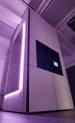
TFS Spectra 300

The TFS Spectra 300 is a state-of-the-art FEG Scanning Transmission Electron Microscope (S/TEM) with a high-tension voltage range of 30 kV - 300 kV. It is completely enclosed in a casing, specially designed to dampen acoustic and temperature variations from the environment. This enclosure not only makes it possible to transfer information well below 1 Å, it also allows the system to easily reach ultra-high resolution routinely in a noisier environment. With Spectra 300 is equipped with a high-brightness X-Feg monochromated source, a piezo-enhanced CompuStage, and two Cs correction optics. The S-CORR above the (S-TWIN) objective lens is used to form electrons probes with sub-Ångström and a CETCOR below the objective lens can be used for high-resolution TEM imaging with a bottom-mounted, retractable, fast Ceta CMOS camera. The Spectra is equipped with a multitude of detectors that allow us to perform multi-model imaging techniques. A Super-X detector with effectively 0.7 srad collection angle allows elemental mapping by secondary X-ray emissions with high resolution. The column is also equipped with a Gatan Continuum 1066 energy filter (GIF) for energy-filtered imaging and spectroscopy with well below 1 eV energy resolution. STEM imaging can be performed with a multichannel segmented Panther detector, a pixelated EMPAD detector, bright-field and dark-field detectors of the GIF, and a standard Fischione dark-field detector. Optical alignment are available for the beam energies 30 keV, 60 keV, 200 keV, and 300 keV.
It is mandatory to store all data recorded with the Spectra microscope using the ER-C Data Management and to generate an Electronic Laboratory Notebook for all measurements.
Storing data on the microscope PC is only allowed temporarily during a session. Please, delete the data after it has been stored in the ER-C Data Management or elsewhere.
Users of the Spectra 300 are kindly asked to quote a technical description of the instrument published in the [tba] when referring to the use of this instrument in publications.
Status
Status, issues and issue reporting: Spectra issue board
Chat for discussion of issues: iffchat-Spectra
Applications
The configuration of the Spectra allows a variety of advanced transmission electron microscopy techniques to be applied to a wide range of solid state materials. These techniques include
- high-resolution coherent TEM imaging
- high-resolution STEM imaging with a multitude of detectors
- high-resolution STEM diffraction imaging (4D-STEM)
- elemental mapping using electron energy loss spectroscopy (EELS) and energy-dispersive X-ray spectroscopy (EDX)
- monochromated EELS
- precession diffraction
- electron tomography
- focal series reconstruction by TrueImage Professional
- automated measurement protocols via TEM scripting API
The Spectra 300 is not intended for the investigation of aqueous, contaminated, ferro-magnetic or organic samples without further discussions with the instrument officers and the ER-C general management. We would also recommend to focus on high-resolution applications and those requiring better EDX sensitivity. Please, use other facilities of the Ernst Ruska-Centre for low resolution, standard imaging and work with high likelihood of contamination.
Access
We are targeting for a high external user project usage and would therefore give priority to those experiments. Also, try to minimize the number of changes of the accelerating voltage.
Instructions
The operation instructions given below and on the subsequent pages are ment as guidelines and knowledge base. They do not replace the direct introduction to the microscope usage by your project manager or the instrument officers. There are many steps and pitfalls in the operation of this microscope that can seriously harm you or the expensive equipment.
Since the Spectra 300 is a very complex machine, please do not edit the instructions below before discussing the changes with the instrument officers. These instructions are also for internal use only, the information should not be published outside the Forschungszentrum Jülich.
Microscope operation
★ Refill the liquid nitrogen dewar when it shows less than 50 % fill state! The microscope uses more than 30 % of the dewar per day. ★
- General instructions
- Monochromator tuning procedures
- STEM (S-CORR) tuning and Diffraction
- TEM (CETCORPlus) tuning
- Working with Velox
- Imaging with the Ceta camera
- STEM imaging
- EDX - tuning procedures
- EELS - Continuum GIF tuning procedures
- Spicer active magnetic field compensation
- Warm-up, Shutdown and Start
Knowledge
Specifications
Microscope performance
Values given here are provisional and takan from the manufacturer specifications as is.
- Acceleration Voltage: 30 kV, 60 kV, 200 kV, and 300 kV
- Information Limit (TEM)
- @ 300 kV < 0.6 Å (monochromated)
- Resolution (STEM)
- @ 300 kV < 0.5 Å
- System Energy Resolution
- @ 60 kV < 0.03 eV
Note that the above values are as specified on delivery. They may change over time, usually with a tendency of degradation.
Detectors
- CETA 16 Megapixel high speed CMOS camera.
- Super-X EDS Detector
- EMPAD: pixelated STEM detector
- Panther: Multi-Channel segmented STEM detector
- Gatan Continuum 1066 energy filter (GIF) with a scintillator-based CMOS camera.
- Fischione Model 3000 HAADF STEM detector.
Specimen holders
- 3mm low-background double-tilt holder (α range ± 70°, ß range ± 30°), hex-ring mount
- 3mm high-visibility low-background double-tilt holder (α range ± 70°, ß range ± 30°) for EDX, clip mount
- Fischione single-tilt tomography holder
IT
- gateway name: iff142
- detector servers (Ceta, GIF, EMPAD) are connected to the microscope PC in a local network
- move data to the gateway from the microscope PC, then from the gateway to a storage
- offload of EMPAD data is currently only possible via USB drives
- ER-C Data Management
- Electronic Laboratory Notebook
Instrument officers
When opening a service call, refer to tool number D3809.
The Spectra 300 instrument officers are:
Location
Building: 05.2W
Operator room: tba
Microscope room: 2087a
Operator Phone: +49 2461 61 tba